肺炎
肺炎,新年给我的见面礼。
放疗中后期,咳嗽加重,少量白痰。时有低烧。
放疗科医生说可能有肺炎,由放疗所致,不是感染,无需治疗。
1月13日,PET扫描。新冠原因,厂家无法供货,医院缺口服对照液,结果难以和以往扫描严格对比。
报告显示有严重肺内感染。淋巴结的SUV增加,这与预期的放疗效果相反,唉……
1月20日,原定化疗日。
肿瘤医生仔细观察PET成像,并与前几次反复对比,认为:
1. 左肺内有大面积严重感染,必须立即用阿奇霉素抗菌,加上强地松疗法抗炎。两周复查。
2. 暂停Alimta化疗。
3. 目前无法评估放疗效果,需要跟踪观察两个月,至少等肺内感染得到控制,才能做准确的判断。
去取了药,并接受长达五分钟的prednisone(强地松)激素疗法用药指导。这个疗法比较麻烦,从每天四片(80mg)起,每三天减半片,直到0片。
至今(2023-02-01),一周的抗菌己结束。咳嗽减轻,基本上没有痰了。
为期三周多的激素减量疗法仍在继续。
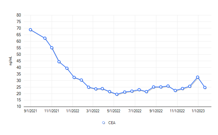
唯一的好消息是,CEA降了。也许新年真有新气相

2023-02-03 复诊。只验了血,加查体
中性粒明显升高,提示有细菌感染。红细胞己恢复正常限。肝、肾功能正常。
约了十天后做CT, 两周复诊。
附
2023-01-13 PET report
Impression
1. Interval development of markedly hypermetabolic diffuse
peribronchial and interstitial opacities. Stable to minimally
increased mildly hypermetabolic pleural thickening. Findings may be
seen in the setting of postradiation inflammatory sequelae. Continued
imaging follow-up recommended to evaluate for underlying infection
versus neoplastic process.
2. Persistent mildly FDG avid aorto esophageal lymph node and
increasing FDG avidity of a morphologically stable left hilar lymph
node.
3. Increased FDG avidity in the left anterior hemithorax corresponding
to sclerotic left rib lesion, SUV 3.0 (prior SUV 2.0 with possible
adjacent pleural involvement.
4. Stable moderate left pleural effusion and increased small right
pleural effusion.
上一篇:https://blog.wenxuecity.com/myblog/73054/202212/33680.html