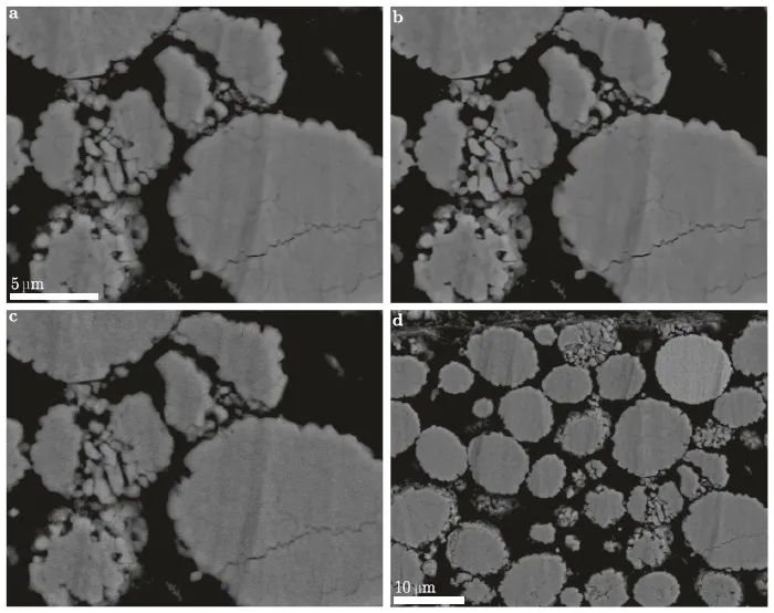
在材料科学领域,显微图像数据的超分辨率解析引起了研究人员极大的兴趣,因为成像技术通常很耗时,并且在材料的成像面积/体积与所达到的分辨率之间存在此消彼长的关系。更准确地说,以小像素/体素尺寸(即高分辨率)执行的成像可以捕获材料微观结构的更多细节,然而,这会导致被成像材料的面积/体积更小。因此,由于局部材料的异质性,通过高分辨率成像获得的单张图像可能不具有统计代表性。另一方面,低分辨率成像可以捕获更大的区域/体积,但可能看不到微观结构的精细细节。这种视野和分辨率相互制约的困境在锂离子电池领域尤为普遍,其中电极具有多尺度结构异质性,每个都需要分析代表性区域以进行准确表征。例如,电极颗粒具有形状和大小的分布,需要足够大的视野来捕获大量颗粒,从而提供其形态的代表性表征。对于在相对较大的体积上变化很大的极小特征,需要大视场和高分辨率。这在 SEM 的情况下既耗时又昂贵。
来自德国乌尔姆大学的Furat等,使用略微修改的生成式对抗网络 (GAN),即SRGAN,对锂离子电池正极内不同老化的 LiNi1−x−yMnyCoxO2(NMC) 颗粒的 SEM 图像数据进行了超分辨率解析。为了训练 SRGAN,他们使用成对的实验测量的低分辨率 SEM 图像和相应的(实验测量的)高分辨率图像,并将 SRGAN 获得的超分辨率结果与他人的获得的超分辨率显微镜图像数据进行定量比较,发现经过训练的 SRGAN解析的 NMC 粒子超分辨率SEM 图像,优于迄今为止文献中的网络方法解析的图像。另外,他们还研究了在没有低分辨率和高分辨率的图像对可用的情况下SRGAN的超分辨能力,发现它可以可靠地提高实验测量图像数据的分辨率,以获得更详细但具有统计代表性的显微镜图像数据。这一方法不限于正极材料的 SEM 图像数据,还可以很容易地推广到通过不同测量技术(如,原子力显微镜)获得的图像数据上。该文近期发表于npj Computational Materials 8:68 (2022),英文标题与摘要如下,点击左下角“阅读原文”可以自由获取论文PDF。
Super-resolving microscopy images of Li-ion electrodes for fine-feature quantification using generative adversarial networks
Orkun Furat, Donal P. Finegan, Zhenzhen Yang, Tom Kirstein, Kandler Smith & Volker Schmidt
For a deeper understanding of the functional behavior of energy materials, it is necessary to investigate their microstructure, e.g., via imaging techniques like scanning electron microscopy (SEM). However, active materials are often heterogeneous, necessitating quantification of features over large volumes to achieve representativity which often requires reduced resolution for large fields of view. Cracks within Li-ion electrode particles are an example of fine features, representative quantification of which requires large volumes of tens of particles. To overcome the trade-off between the imaged volume of the material and the resolution achieved, we deploy generative adversarial networks (GAN), namely SRGANs, to super-resolve SEM images of cracked cathode materials. A quantitative analysis indicates that SRGANs outperform various other networks for crack detection within aged cathode particles. This makes GANs viable for performing super-resolution on microscopy images for mitigating the trade-off between resolution and field of view, thus enabling representative quantification of fine features.